[最も人気のある!] paranasal sinuses easy diagram 130211
Media in category "Paranasal sinuses" The following 35 files are in this category, out of 35 total 3D printed upper respiratory airways cast jpg 2,560 × 1,9;Humans possess four paired paranasal sinuses, divided into subgroups that are named according to the bones within which the sinuses lie The maxillary sinuses, the largest of the paranasal sinuses, are under the eyes, in the maxillary bones (open in the back of the semilunar hiatus of the nose) They are innervated by the trigeminal nerve (CN V2)Paranasal Sinuses STUDY Learn Write Test PLAY Match − Created by jace_robinson5 PLUS Terms in this set (4) Frontal Sinus Sphenoidal Sinuses Maxillary Sinuses Ethmoidal Labyrinth (Sinuses) THIS SET IS OFTEN IN FOLDERS WITH Nasal Conchae 7 terms jace_robinson5 PLUS Skull Cranial Cavity 13 terms jace_robinson5 PLUS Mandible 10 terms jace_robinson5
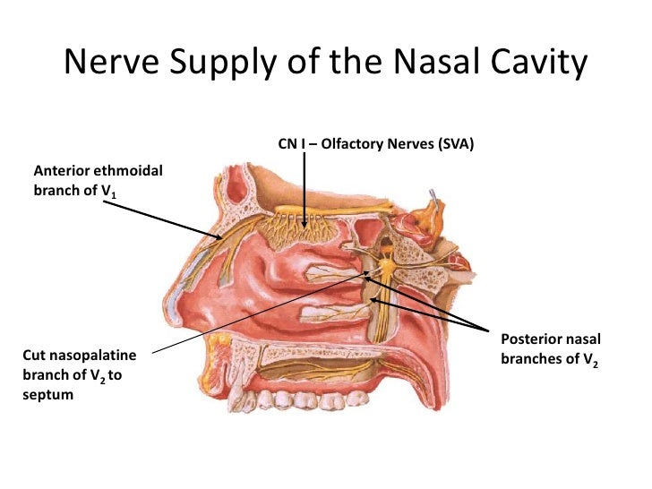
Anatomy Of Nasal Cavity Anatomy Drawing Diagram
Paranasal sinuses easy diagram
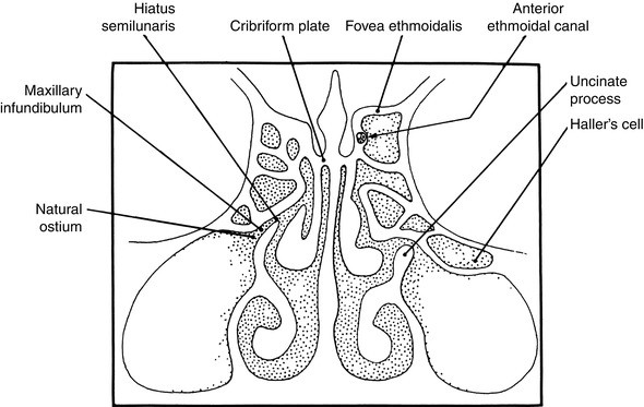
Paranasal sinuses easy diagram-Anatomic variations of the nasal cavity and paranasal sinuses are described in order of their appearance in endoscopy Variations of the pyriform aperture are followed by variations of the agger nasi, inferior and middle conchas, inferior and middle nasal meati, and variations of the bulla ethmoidea, uncinate process and fontanelles Variations of the maxillary sinus are describedFor a righthanded person the conflict is associated with his/her mother or child HEALING PHASE During the first part of the healing phase (PCLA) the tissue loss is replenished through cell proliferation
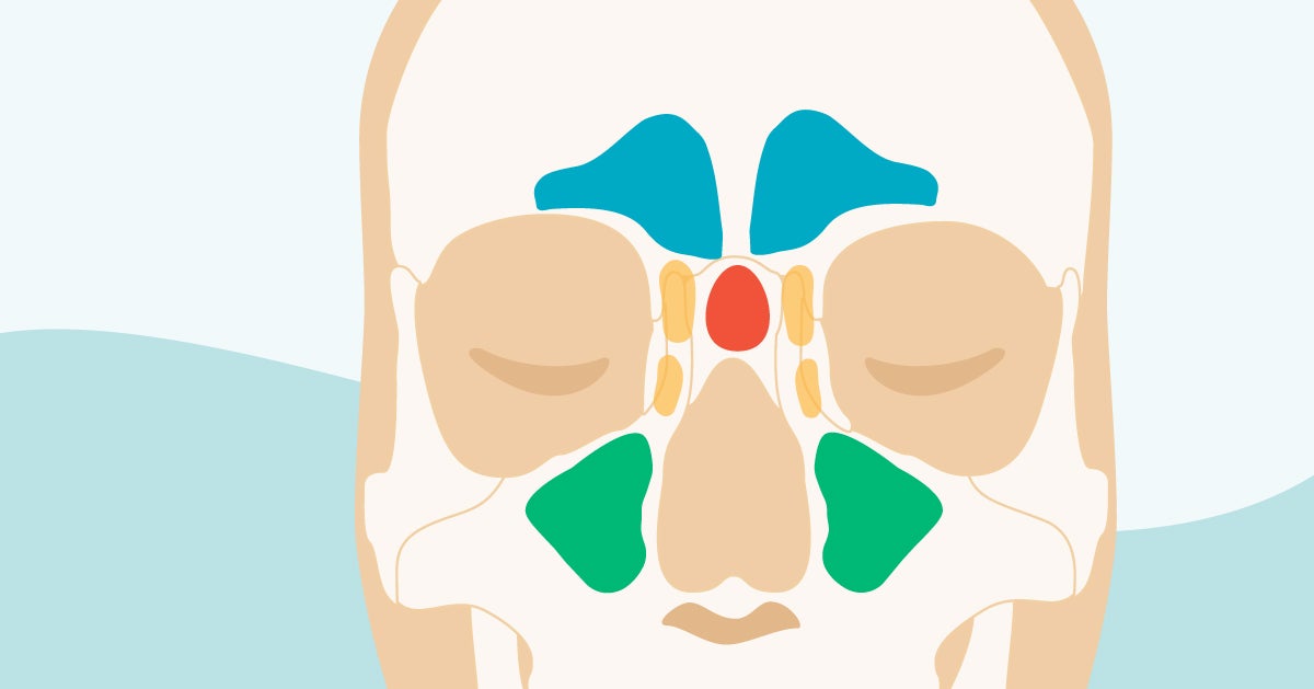


Sinus Cavities In The Head Anatomy Diagram Pictures
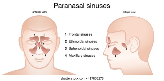
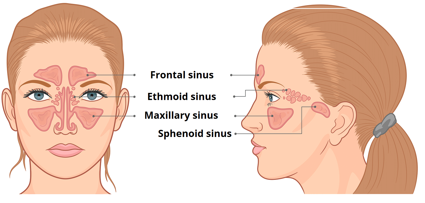
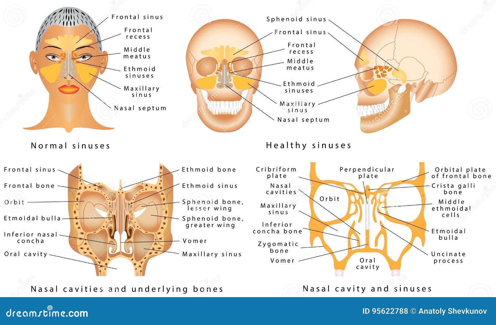
May 14, 13 Nasal sinus and paranasal sinus are part of nasal anatomy frontal sinus cavity, nasal sinus, nasal bone are here with imagesThere are four groups of paranasal sinuses the maxillary, ethmoid, frontal, and sphenoid sinuses (see figure Locating the Sinuses) Sinuses reduce the weight of the facial bones and skull while maintaining bone strength and shape The airfilled spaces of the nose and sinuses also add resonance to the voice Locating the Sinuses The supporting structure of the upper part of theParanasal sinuses refer to a group of airfilled spaces around the nasal cavity (a system of air channels that connect the nose with the back of the throat) (1) They facilitate the circulation of the air breathed in and out of the respiratory system (2) Paranasal sinuses have four different pairs maxillary sinuses, frontal sinuses, sphenoidal sinuses, and ethmoidal sinuses (3) Their names
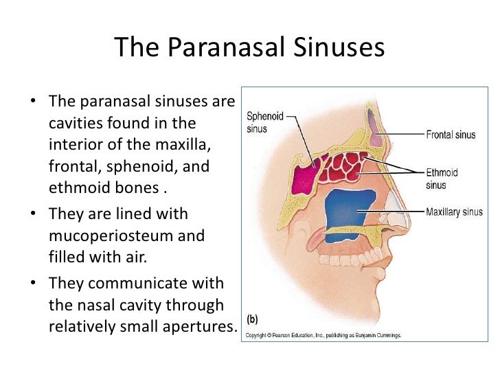
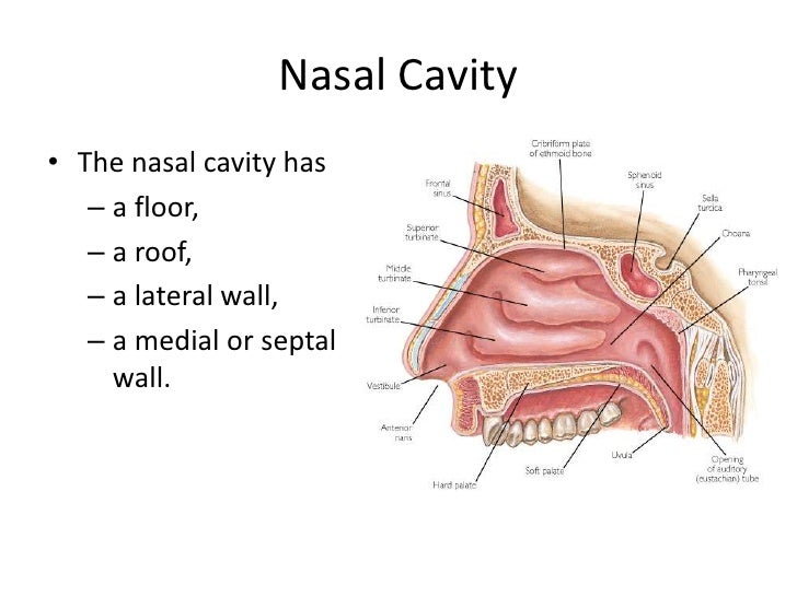
Fig 222 Schematic drawing of paranasal sinuses, showing AP relationship to each other and surrounding parts Intersinus septum Sphenoidal sinuses Maxillary sinuses Frontal sinuses Ethmoidal sinuses Fig 221 Anterior aspect of paranasal sinuses, showing lateral re lationship to each other and to surrounding parts Fig 225 Underexposed radiograph of sinuses g 224 · ANATOMY AND PHYSIOLOGY OF PARANASAL SINUSES NAVEENK 2 Air filled spaces present within certain bones of the skull, around the nasal cavities "PARANASAL SINUSES" 3 FRONTAL SINUS Location In the frontal bone, above and deep to the supra orbital margin Average measurements ht 32cm, width 24cm, depth 16cm Usually asymmetricalThe paranasal sinuses The paranasal sinuses are airfilled cavities in the frontal, ethmoid, maxilla, and sphenoid bones They're lined with a mucosal membrane and have small openings into the nasal cavity Maxillary sinus This sinus is located in the body of the maxilla behind the cheek just above the roots of the premolar and molar teeth It's shaped like a pyramid It opens into the
The tongue has two parts (see diagram above) The front part is the part you can see and it makes up twothirds of the tongue The back part, which you cannot see, is very close to the throat We have more information about tongue cancer The throat (pharynx) The throat is divided into three main parts Nasopharynx This is the upper part of the pharynx behind the nose Cancer thatFrontal sinus, sphenoidal sinus,; · The paranasal sinuses are airfilled cavities within the ethmoid, frontal, and sphenoid bones and within the maxilla (Fig 175) They serve as resonating chambers for the voice and help to warm and moisten inhaled air The sinuses develop during childhood and are not fully formed until age 16 to 18 At maturity, passages connect the sinuses to each other and to the nasal cavity
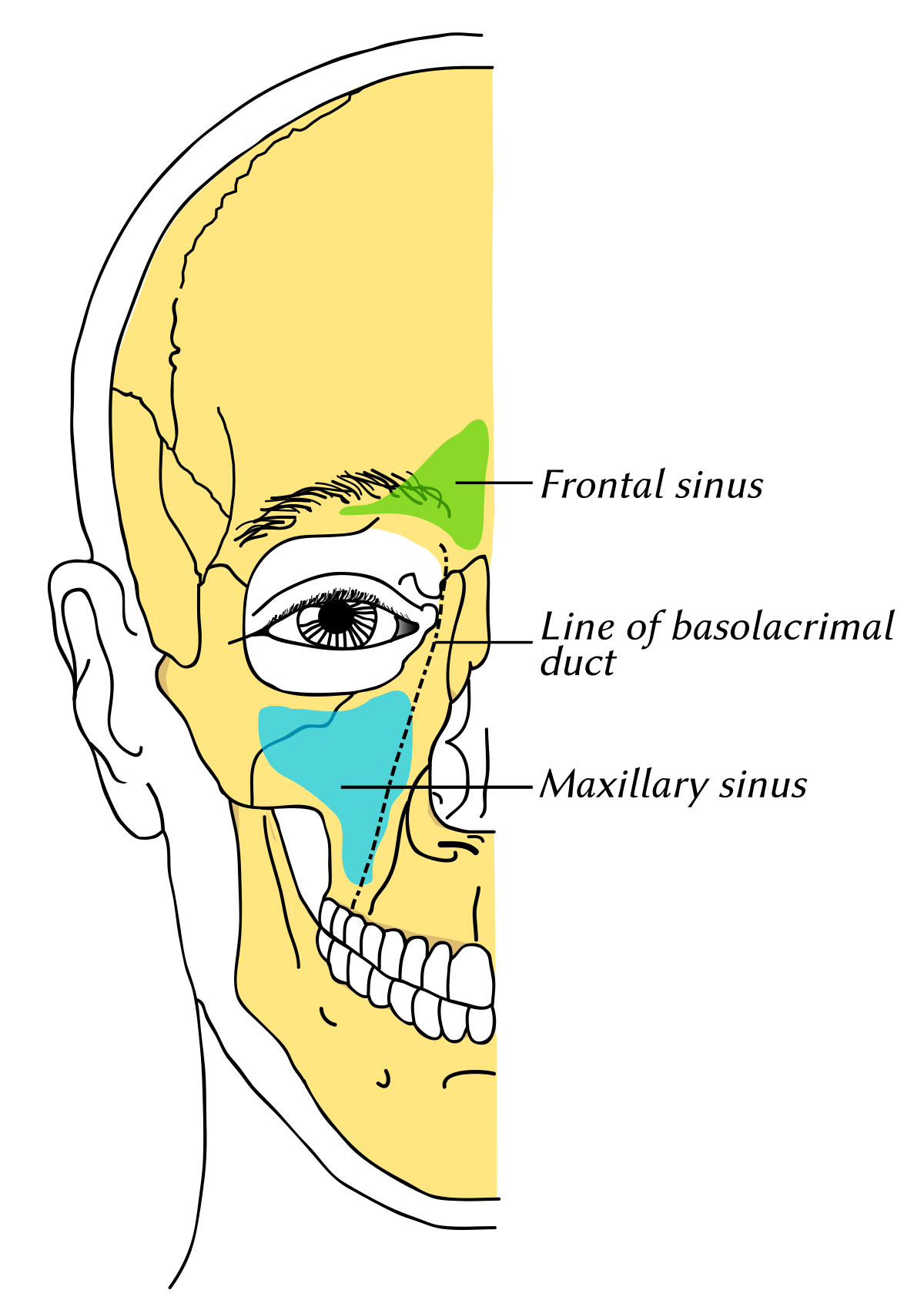


Maxillary Sinus Wikipedia
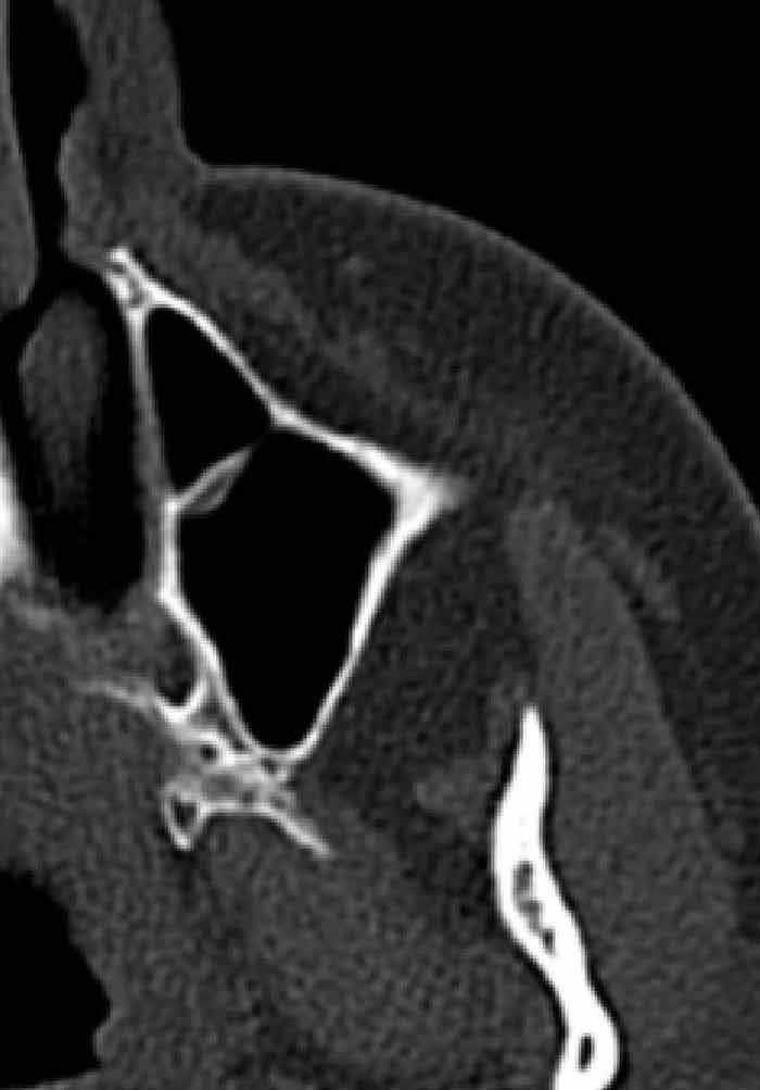


Paranasal Sinus Anatomy What The Surgeon Needs To Know Intechopen
· These sinuses collectively are called the paranasal sinuses The name sinus comes from the Latin word sinus , which means a bay, a curve, or a hollow cavity Picture of the sinusesNasal Polyps ( Small growth in the lining of the nose ) Allergic rhinitis ( swelling of the lining of · Maxillary air sinuses are the largest paranasal sinuses Location Are located in the body of maxilla lateral to the nasal cavity and inferior to the floor of orbit Dimensions Capacity15ml Height 35cm, Anterior posterior depth – 35cm, Width 25cm Boundaries and relations Maxillary air sinuses are pyramidal in shape and have


21 Best Paranasal Sinuses Ideas Paranasal Sinuses Sinusitis Radiology


Paranasal Sinuses Maxillary Ethmoid Sphenoid Frontal Notes Youtube
· Paranasal sinuses are air filled hollow sacs seen around the skull bone These sacs precisely surround the nasal cavity There are four paired sinuses surrounding the nasal cavity · The maxillary sinuses are the largest of the all the paranasal sinuses They have thin walls which are often penetrated by the long roots of the posterior maxillary teeth The superior border of this sinus is the bony orbit, the inferior is the maxillary alveolar bone and corresponding tooth roots, the medial border is made up of the nasal cavity and the lateral and anterior borderAnatomy and physiology of the nose and paranasal sinuses Immunol Allergy Clin N Am 04; Warner N, Eggenberger E Traumatic optic neuropathy a review of the current literature Curr Opin Ophthalmol 10; Watters EC, et al Acute orbital cellulitis Arch Ophthalmol 1976;4785 Weber AL, Mikulis DK Inflammatory disorders of the paraorbital sinuses and their


Sinus Cavities In The Head Anatomy Diagram Pictures
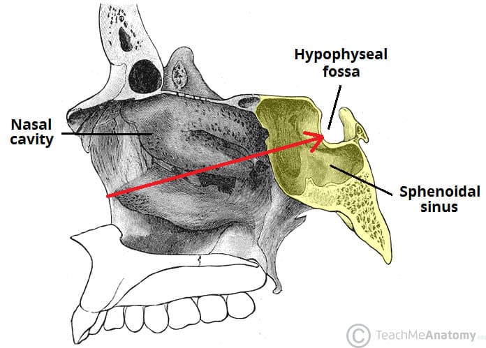


The Paranasal Sinuses Structure Function Teachmeanatomy
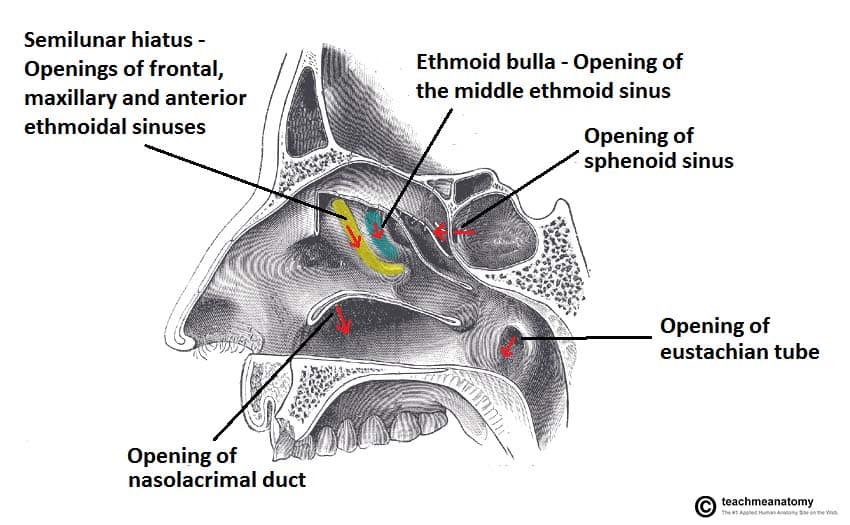
Endoscopes are used to restore drainage of the paranasal sinuses and ventilation of the nasal cavity Brief information regarding this surgery has been offered Functional endoscopic sinus surgery is a popular surgery used for treating sinus infections It is meant for treating sinusitis and nasal polyps, bacterial, fungal, recurrent acute, chronic sinus problems Endoscopes are used to · Paranasal sinuses are airfilled spaces present within some bones around the nasal cavities The sinuses are Frontal, maxillary, sphenoidal, and ethmoidal All of them open into the nasal cavity through its lateral wall The function of the sinuses is to mack the skull lighter and add resonance to the voice · WebMD's Sinuses Anatomy Page provides a detailed image and definition of the sinuses including their function and location Also learn about conditions, tests, and treatments affecting the sinuses
:background_color(FFFFFF):format(jpeg)/images/article/en/the-paranasal-sinuses/972PC0nYOzlz7wqSgLmNA_sinus_frontalis_large_u9Vfozc0uUoMtc6KtIaUfw.png)


Paranasal Sinuses Anatomy And Clinical Aspects Kenhub


Assessment Of Human Sinus Cavity Air Volume Using Tunable Diode Laser Spectroscopy With Application To Sinusitis Diagnostics Huang 15 Journal Of Biophotonics Wiley Online Library
Used as a passage for air and other gases to travel · The paranasal sinuses are group of air filled spaces surrounding the nasal cavity Paranasal sinuses start developing from the primitive choana at 25–28 weeks of gestation Three projections arise from the lateral wall of the nose and serve as the beginning of the development of the paranasal sinusesParanasal sinus disease is common and on occasion can become lifethreatening if not treated in a timely fashion At birth the maxillary sinuses and ethmoid air cells are present but hypoplastic The sphenoid sinus develops around 4 years of age secondary to pneumatization of the sphenoid bone The frontal sinuses are the last to pneumatize at around 8–10 years of age so that in theory


Paranasal Sinuses Diagram Quizlet


Amazon Com Paranasal Sinus Watercolor Art Print Anatomy Wall Decor Sinuses Artworks Frontal Section Of The Nasal Cavity Wall Art Sinuses And Sinusitis Office Decor Sinus Cavity Diagram Ent And Omfs Art Handmade
Nasal and sinus cancer affects the nasal cavity (the space behind your nose) and the sinuses (small airfilled cavities inside your nose, cheekbones and forehead) It's a rare type of cancer that most often affects men aged over 40 Nasal and sinus cancer is different from cancer of the area where the nose and throat connect This is called nasopharyngeal cancer Credit InformationOur paranasal sinuses come in pairs, with one of each pair located on either side of the face You have four of these pairs Sinus Anatomy – Paranasal Sinuses Maxillary Sinuses These are found directly in behind the check Ethmoid Sinuses These are smaller pockets of air, often described as having the appearance of honeycombChart and Diagram Slides for PowerPoint Beautifully designed chart and diagram s for PowerPoint with visually stunning graphics and animation effects Our new CrystalGraphics Chart and Diagram Slides for PowerPoint is a collection of over 1000 impressively designed datadriven chart and editable diagram s guaranteed to impress any audience They are all artistically


Diagram Of Paranasal Sinuses Download Scientific Diagram
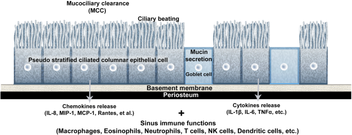


Definition And Management Of Odontogenic Maxillary Sinusitis Maxillofacial Plastic And Reconstructive Surgery Full Text
READ MORE BELOW!In this video, we explore the 4 paranasal sinuses and what their proposed functions areINSTAGRAM @thecatalystuniversityI'm starting an InsApr 29, 16 Explore Natalie StickneyShade's board "paranasal sinuses", followed by 140 people on See more ideas about paranasal sinuses, sinusitis, radiologyNov 29, 16 This Pin was discovered by Alecia Fidler Discover (and save!) your own Pins on
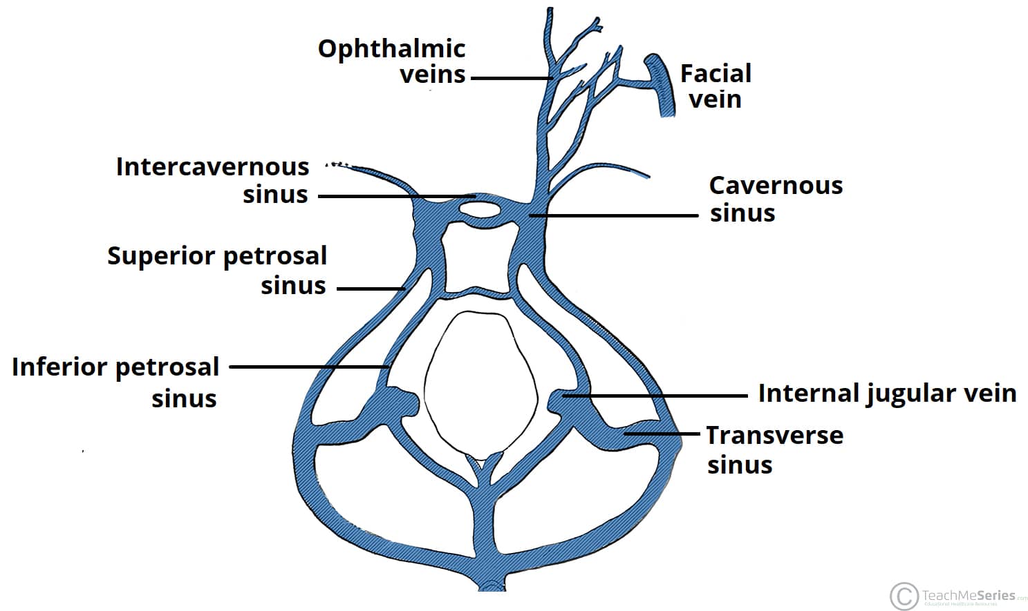


The Cavernous Sinus Contents Borders Thrombosis Teachmeanatomy


Equine Diseases Of Nasal Cavity And Paranasal Sinuses Diagram Quizlet
Paranasal sinuses Dr Sachintha Hapugoda and Dr Maxime StAmant et al The paranasal sinuses usually consist of four paired airfilled spaces They have several functions of which reducing the weight of the head is the most important Other functions are air humidification and aiding in voice resonance They are named for the facial bones inThe right side of the premotorsensory cortex (view the GNM diagram) for the left paranasal sinuses, linked to a scent or stink conflict related to a partner if the person is lefthanded;The paranasal sinuses (latin sinus paranasales) are four bilateral airfilled spaces within bones of the skull surrounding the nasal cavityFour bones of the skull each accommodates a pair of paranasal sinuses that are named according to the bone in which they are locatedThe four sinuses are maxillary sinus,;


Pin On Speech Lang Path


Equine Diseases Of Nasal Cavity And Paranasal Sinuses Diagram Quizlet
· Anatomy of the paranasal sinuses The paranasal sinuses comprise four pairs of sinuses that surround the nose and drain into the nasal cavity by way of narrow channels called ostia (singular ostium) Mucus leaving the frontal (forehead) and maxillary (cheek) sinuses drains through the ethmoid sinuses (behind the bridge of the nose), so a backup in the ethmoids isSinus congestion often seems to be accompanied by excess pressure on the outer ears (as anyone who has shared a transcontinental flight with an infant can attest to) or fluid buildup in the canals An increase the capillary pressure within the ear canals would explain the pressure, but what is the physiological connection between the sinus pressure and pressure in the ears?Sinuses are a hollow space inside the nasal bone filled with air paranasal sinus is called sinusitis A healthy sinus filled with the air but when they filled and blocked with liquid, mucus, germs so it can cause the sinuses infection The conditions that can cause Sinus blockage Common Cold;
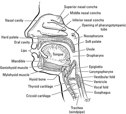


The Anatomy Of The Nose Dummies


Paranasal Sinuses Diagram Quizlet
Learn more Royaltyfree stock vector ID Sinuses of Nose Human Anatomy Sinus Diagram Anatomy of the Nose Nasal cavity bones Anatomy of paranasal sinuses Sinusitis It is the inflammation of the maxillary sinuses l/05/13 · The paranasal sinuses are airfilled extensions of the nasal cavity There are four paired sinuses – named according to the bone in which they are located – maxillary, frontal, sphenoid and ethmoid Each sinus is lined by a ciliated pseudostratified epithelium, interspersed with mucussecreting goblet cellsEthmoid sinus (known as
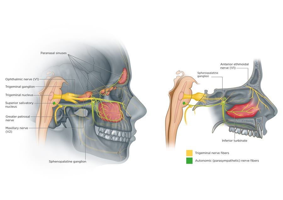


Sinus Migraine A Costly Blindspot In Medical Care Research Outreach
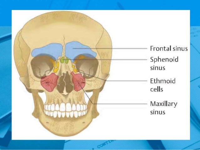


Anatomy And Physiology Paranasal Sinuses
/08/19 · The paranasal sinuses are broadly divided into two major groups the anterior sinuses and the posterior sinuses The anterior sinuses comprise the frontal sinus, anterior ethmoid air cells, and maxillary sinus These drain into a common area centered in the middle meatus, the osteomeatal unit (OMU) The posterior sinuses are the posterior ethmoid air cells and the sphenoid sinuses · Maxillary Sinus (within the maxillary bones) The largest among all the paranasal sinuses 2, these two conical cavities are located on the two sides of the nose, above the upper teeth, and below the cheeks 4 Ethmoid Sinus (within the ethmoid bones) Three to eighteen 5 air cells present in the ethmoid labyrinth, on both sides of the nose, between the eyes 6, 7Paranasal Sinuses STUDY Learn Flashcards Write Spell Test PLAY Match Gravity Created by erika_aranda PLUS Key Concepts Terms in this set (18) Innervation and blood supply to Frontal Sinus Supraorbital Nerve and Artery and Ethmoidal Arteries Frontal Sinus opening into the Nasal Cavity Frontonasal Canal into the Anterior Part of the Middle Meatus in the Luminaris Hiatus


1
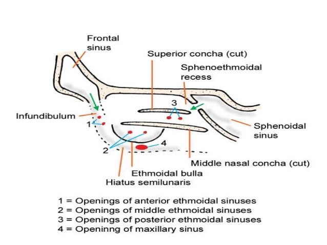


Anatomy Of Paranasal Sinuses
· Anatomically, the paranasal sinuses are in close proximity to the anterior cranial fossa, the cribriform plate, the internal carotid arteries, the cavernous sinuses, the orbits and their contents, and the optic nerves as they exit the orbits 31 – 35 The surgeon should be cautious when maneuvering instruments in the posterior direction to avoid inadvertent penetration and drainage · The paranasal sinuses are airfilled spaces around the nasal cavity that have many possible functions The mucosa of the upper respiratory tract contain antimicrobial proteins that are a barrier component of the innate immune system Key Terms nostril Either of the two orifices located on the nose (or on the beak of a bird);Paranasal sinuses anatomy Paranasal sinuses are air filled cavities in the frontal maxilae ethmoid and sphenoid bones From the nasal mucosa and their continued communication with the nasal fossae The largest sinus cavities are about an inch across 52 6 maxillary sinus see fig What are the paranasal sinuses a formation from They further develop over the first few years of life 11


Nasal Cavity And Paranasal Sinuses Diagram Quizlet


Tumors Of The Nose And Paranasal Sinus Ento Key
151 MB 3D printed upper respiratory airways cast jpg 1,9 × 2,560; · The 4 sets of paranasal sinuses begin their development at the end of the 3rd month of IUL as outpouchings of the mucous membrane of the middle & superior nasal meatus Primary pneumatization The early nasal sinuses expands into the cartilage walls & roof of the nasal fossae by growth of mucous membrane sacs into the respective bones Secondary pneumatization the sinuses


Anatomy Of The Para Nasal Air Sinuses Dr Yusuf Youtube
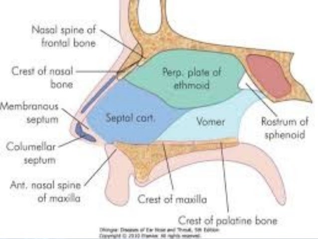


Anatomy And Physiology Of Nose And Paranasal Sinuses


Human Nose Wikipedia
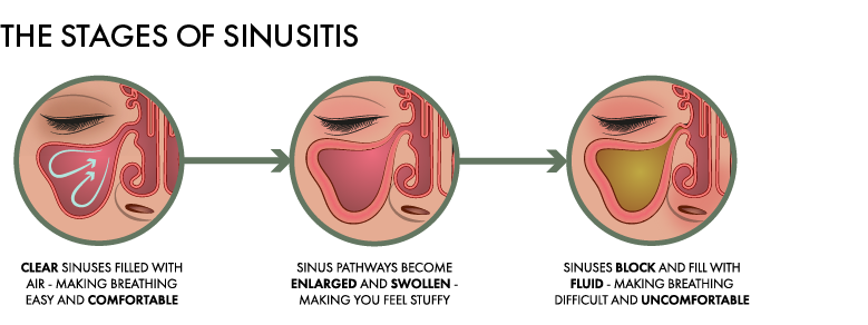


How To Sleep With Sinusitis Sinusitis And Sleep Woolroom
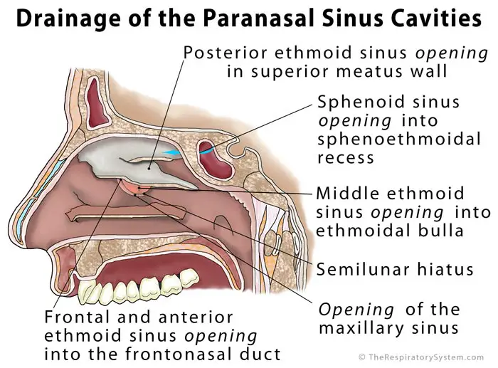


Paranasal Sinus Definition Location Anatomy Function Picture


Models For The Study Of Nasal And Sinus Physiology In Health And Disease A Review Of The Literature Al Sayed 17 Laryngoscope Investigative Otolaryngology Wiley Online Library


Nasal And Paranasal Sinus Anatomy And Embryology Ento Key


Nose And Paranasal Sinuses Anatomy Simplified Youtube
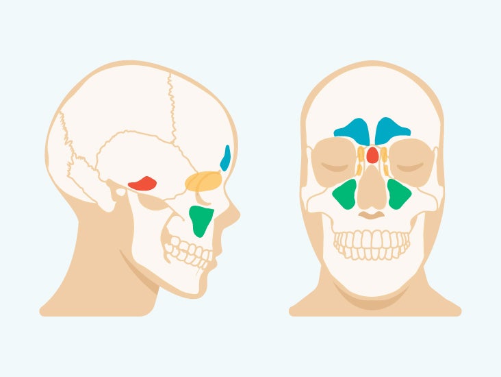


Sinus Cavities In The Head Anatomy Diagram Pictures


Human Nose Stock Illustration Download Image Now Istock
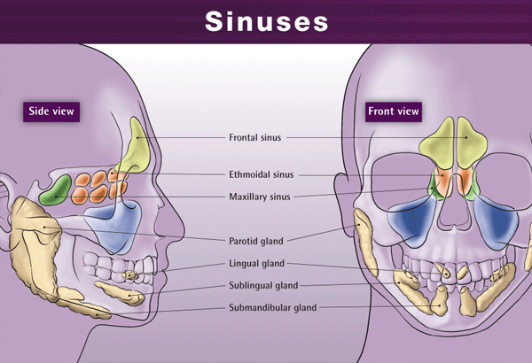


Paranasal Sinuses Everything About Paranasal Sinuses
(107).jpg)


Nose Sinuses Proprofs Quiz


Nose And Paranasal Sinuses Ento Key
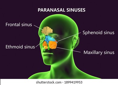


Sphenoid Sinus Images Stock Photos Vectors Shutterstock
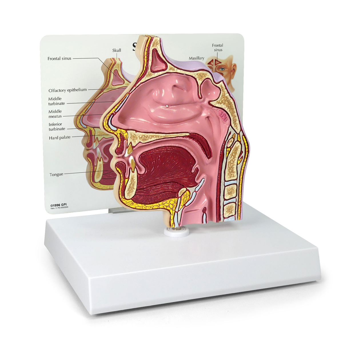


Sinus Cross Section 2850 Anatomical Models Anatomy Teaching Models Cranial Models Head Models
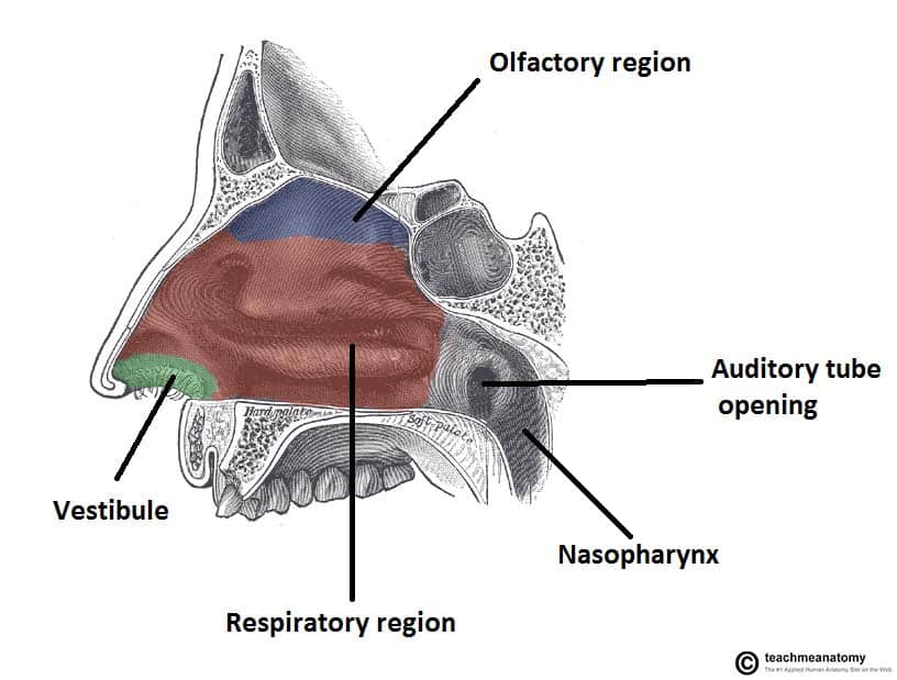


The Nasal Cavity Structure Vasculature Innervation Teachmeanatomy


A Coronal Schematic Representation Of The Posterior Maxillary Teeth Download Scientific Diagram


Emvency Wall Tapestry Sinuses Of Nose Human Anatomy Sinus Diagram The Nasal Cavity Bones Paranasal Sinusitis It Is Inflammation Maxillary Decor Wall Hanging Picnic Bedsheet Blanket 60x50 Inches Amazon Co Uk Home


Maxillary Sinus Anatomy Function Function Body Maps


Sinusitis Animation Youtube


Pin On Teaching Materials
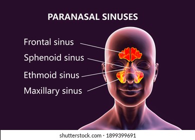


Sphenoid Sinus Images Stock Photos Vectors Shutterstock


1


Pin On Glandular Odontogenic Cysts


Jaypeedigital Ebook Reader


Pin On Health Wellness


Overview Of The Drainage Pathways Of The Paranasal Sinuses A Download Scientific Diagram


Middle Ear And Sinus Problems Skybrary Aviation Safety
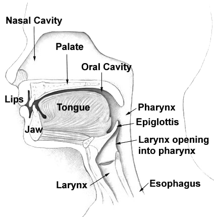


Pharynx Wikipedia


Diagram Of Paranasal Sinuses Download Scientific Diagram


Paranasal Sinuses Diagram Quizlet


Paranasal Sinuses Human Anatomy Organs
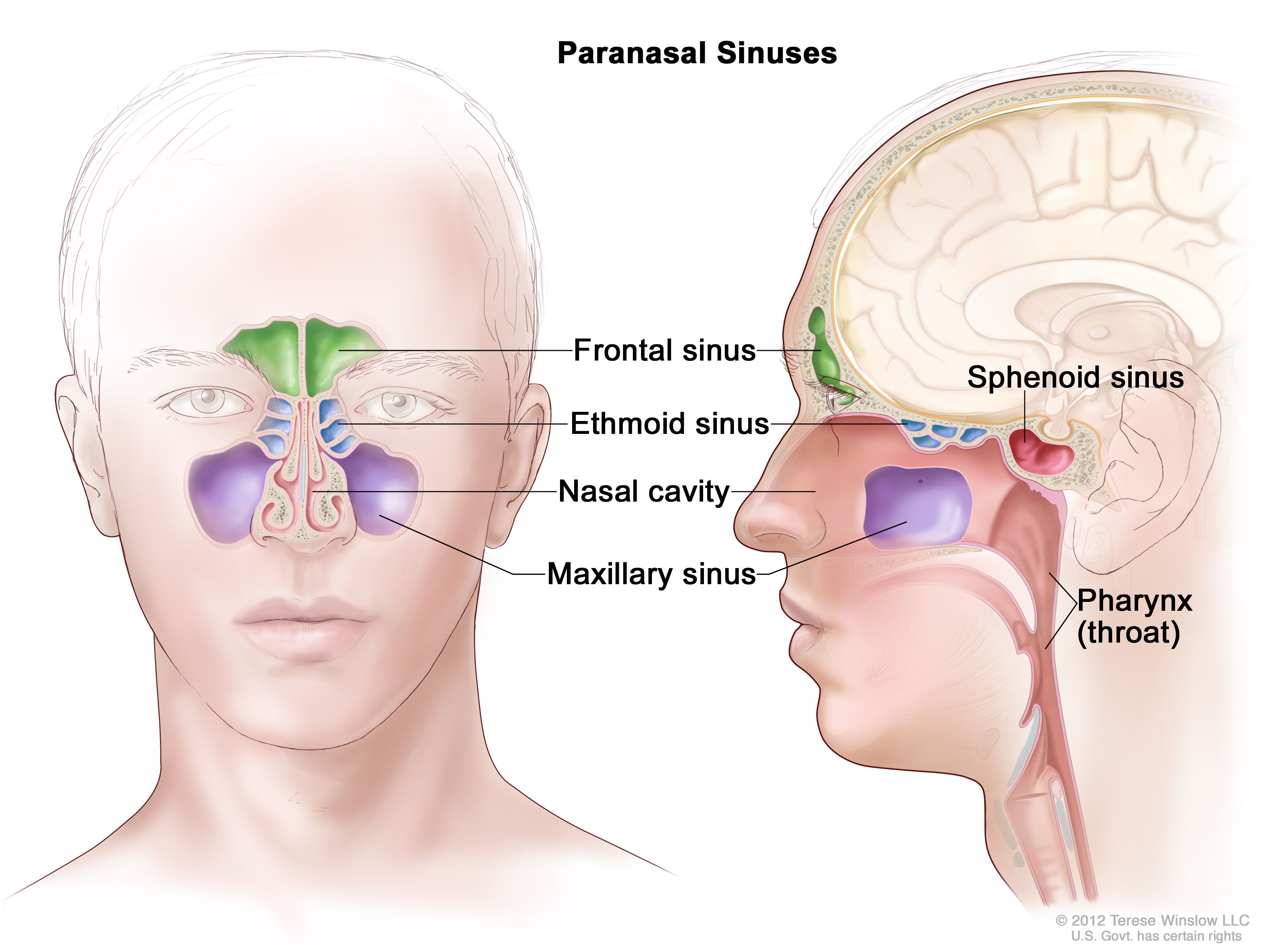


Definition Of Paranasal Sinus Nci Dictionary Of Cancer Terms National Cancer Institute


Diagram Of Paranasal Sinuses Download Scientific Diagram
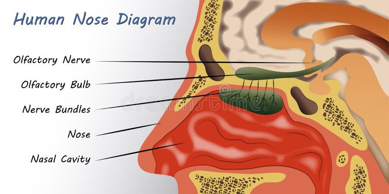


Nose Diagram Stock Illustrations 1 544 Nose Diagram Stock Illustrations Vectors Clipart Dreamstime


Nose Useful Notes On Human Nose And Para Nasal Sinuses Human Anatomy


Schematic Side And Front View Of The Anatomy Of The Human Nasal Cavity Download Scientific Diagram


Paranasal Sinus High Res Illustrations Getty Images


2 8 Paranasal Sinuses Diagram Quizlet


Anatomy Of Nasal Cavity Anatomy Drawing Diagram


Re2 Paranasal Sinuses Diagram Quizlet
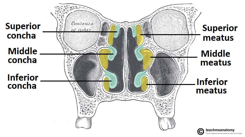


The Nasal Cavity Structure Vasculature Innervation Teachmeanatomy


Maxillary Sinus Cancer Tnm Stages And Grades Cancer Research Uk
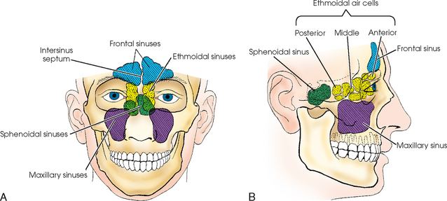


Paranasal Sinuses Radiology Key


Nasal And Paranasal Sinus Anatomy And Embryology Ento Key
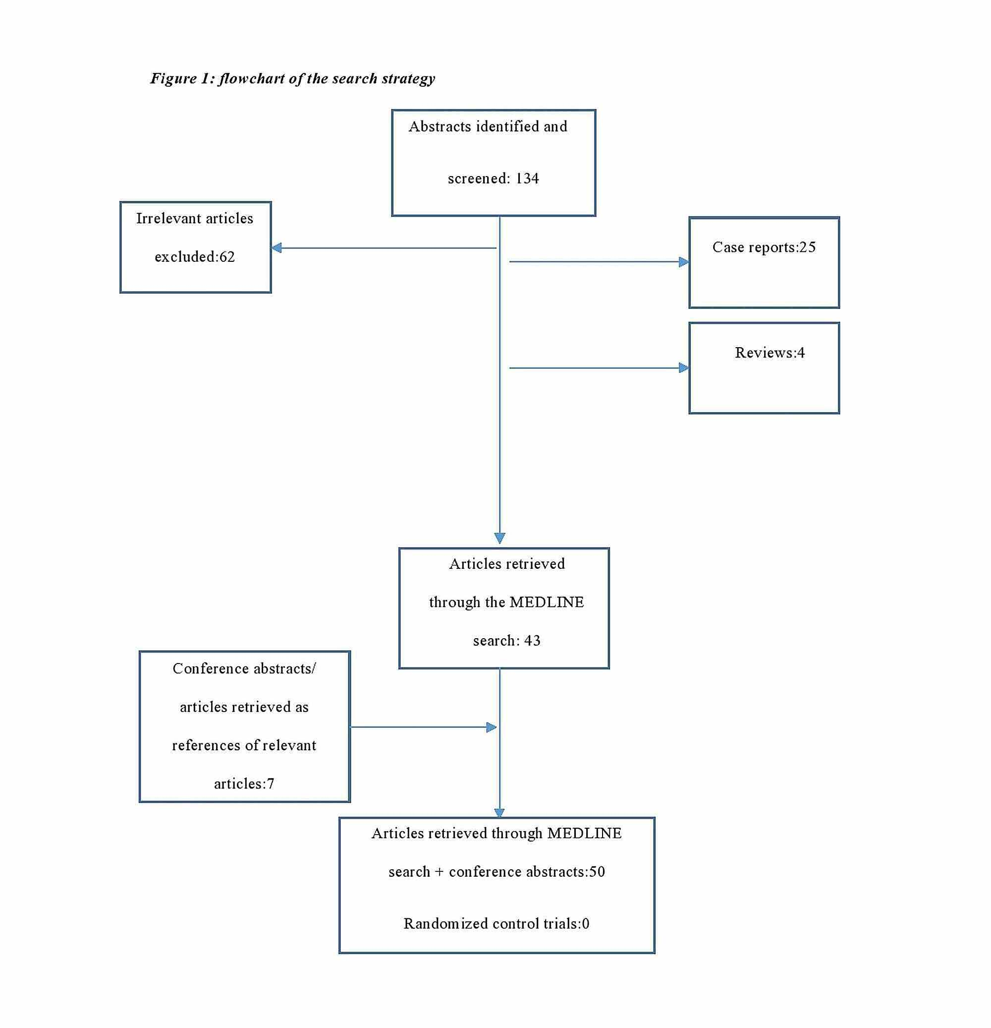


Cureus Anatomical Variations Of The Nasal Cavities And Paranasal Sinuses A Systematic Review
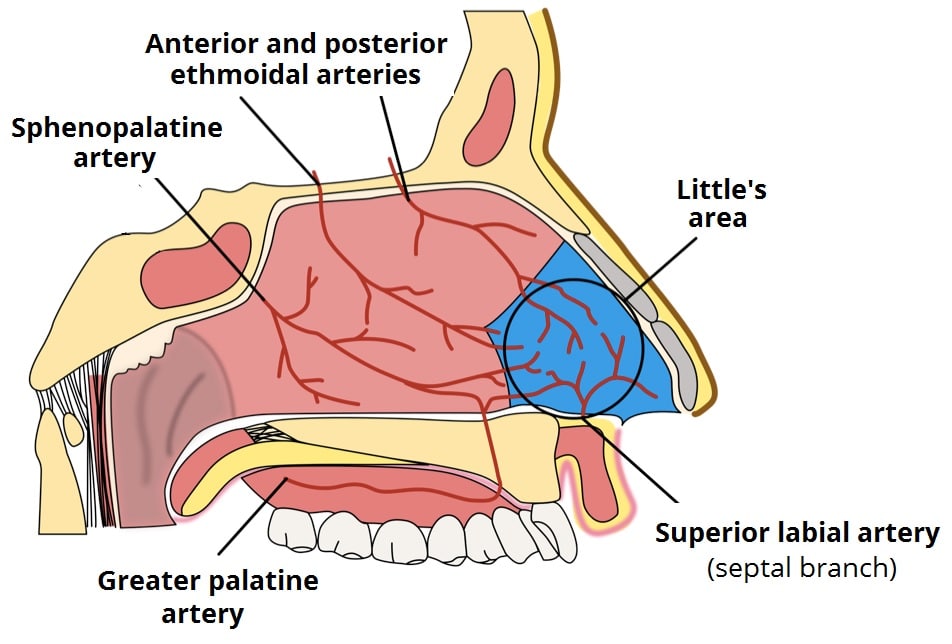


The Nasal Cavity Structure Vasculature Innervation Teachmeanatomy


Pin On Paranasal Sinuses


1


Nose Description Functions Facts Britannica


Sphenoid Sinus Images Stock Photos Vectors Shutterstock


Paranasal Sinus High Res Illustrations Getty Images


What Are Sinuses Anatomy Types Study Com


The Paranasal Sinuses Structure Function Teachmeanatomy


Schematic Representation Of No Production Within Airways The Paranasal Download Scientific Diagram


9 Nasal Cavity Ideas Nasal Cavity Anatomy Sinus Cavities


Nose Diagram Stock Illustrations 1 544 Nose Diagram Stock Illustrations Vectors Clipart Dreamstime


Medicowesome Memorizing How To Draw The Nasal Septum Hey


Paranasal Sinuses Diagram Quizlet


Anatomy Of The Nose And Paranasal Sinuses Springerlink


Middle Ear And Sinus Problems Skybrary Aviation Safety


Paranasal Sinuses Wikipedia


Paranasal Sinuses Diagram Quizlet


Anatomy Of Nose And Paranasal Sinus


Anatomy Of Nose And Paranasal Sinus


Sinus Diagram Stock Illustrations 337 Sinus Diagram Stock Illustrations Vectors Clipart Dreamstime


The Paranasal Sinuses Structure Function Teachmeanatomy
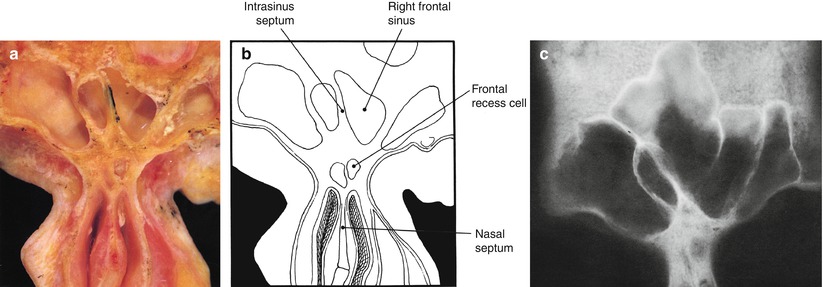


Anatomy Of The Nose And Paranasal Sinuses Springerlink


Nasal And Paranasal Sinus Anatomy And Embryology Ento Key


1
(166).jpg)


Nasal Cavity And Paranasal Sinuses Quiz Proprofs Quiz


21 Best Paranasal Sinuses Ideas Paranasal Sinuses Sinusitis Radiology


Paranasal Sinuses


Draw The Nasal Septum With Me Youtube


What Are Sinuses Anatomy Types Study Com


Diagram Of Paranasal Sinuses Download Scientific Diagram


Paranasal Sinus High Res Illustrations Getty Images


Organs And Structures Of The Respiratory System Anatomy And Physiology


Nasal And Sinus Conditions Of Childhood Michael Rothschild Md
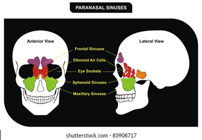


Sphenoid Sinus Images Stock Photos Vectors Shutterstock


コメント
コメントを投稿